General Electric monogramGE Heritage Village
GE Heritage Village bulb logo

GE STEM Lab · Student course

Healthcare & Medical Science

Explain the magnet → radio pulse → signal → reconstructed image sequence without confusing MRI with X-rays.

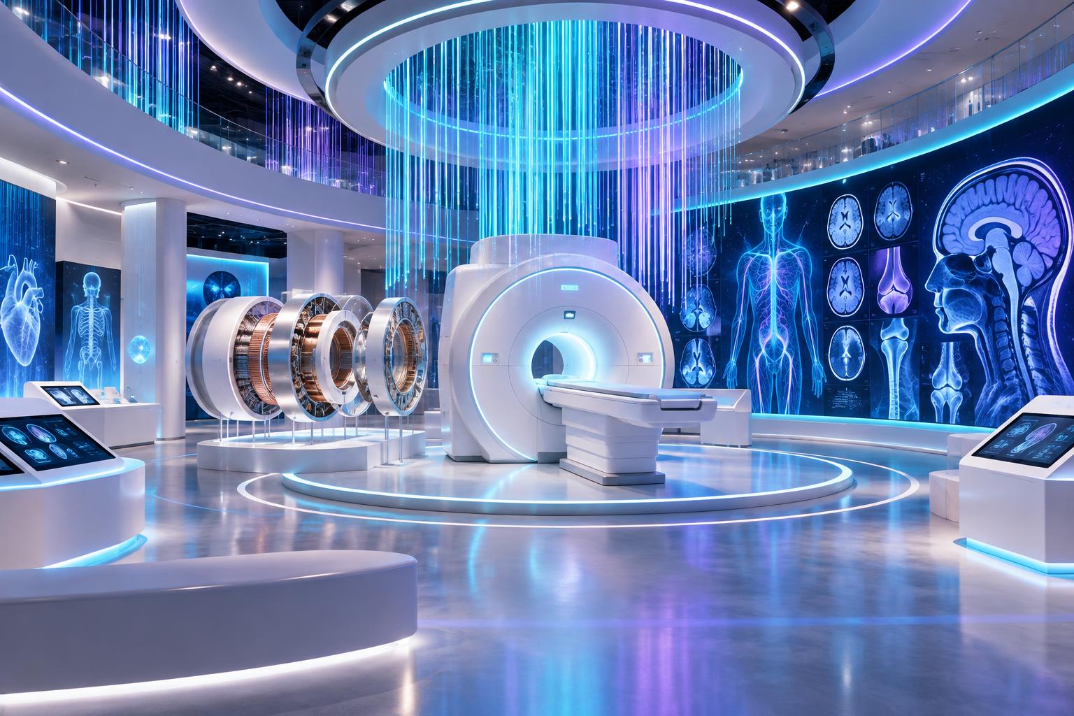
Original imagined learning zone · AI-assisted concept art, not a real GE facility.

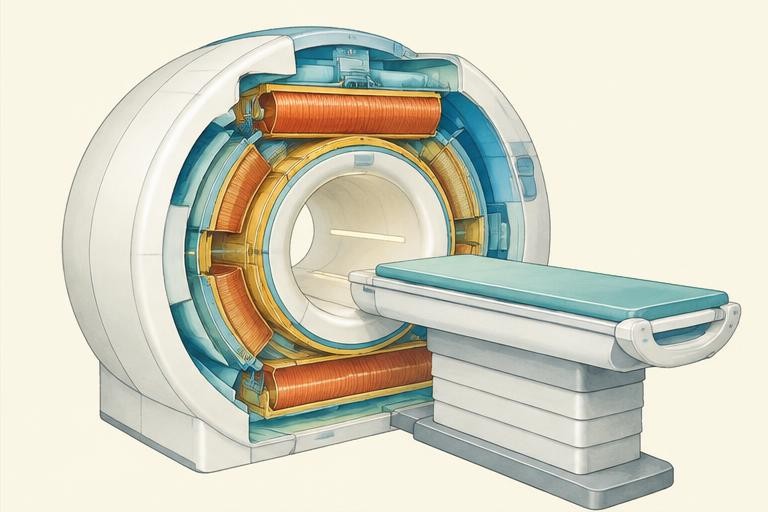
Labeled system diagram · Healthcare & Medical Science
1234Encoded signalsMagnetic field + RF + spatial encoding
  1. 1

    Main magnet

    Creates a strong static magnetic field and a small net nuclear magnetisation.

  2. 2

    RF coil

    Transmits radiofrequency pulses and receives signals.

  3. 3

    Gradient coils

    Vary the magnetic field with position for spatial encoding.

  4. 4

    Reconstruction

    A computer uses encoded signals to construct an image; this is not a real patient scan.

Original simplified teaching schematic—not to scale, an installation guide, a historical artifact or a complete technical blueprint.

🧲 HALL COURSE · 4 COMPLETE LESSONS

Healthcare & Medical Science · learning syllabus

Explain the magnet → radio pulse → signal → reconstructed image sequence without confusing MRI with X-rays. Medical engineering combines physics, electronics and computing to create imaging equipment.

Suggested 100 minutes · foundation explanations → worked examples → interactive experiments

Physics · Waves · 20 suggested minutes

01 · Waves, electromagnetic radiation and imaging

Learning objective: Compare radio waves and X-rays without treating them as the same imaging method.

Waves transfer energy without the bulk transport of matter. Wave speed v = fλ links frequency f and wavelength λ. Electromagnetic waves are transverse and can travel through a vacuum. In a vacuum they share the same speed, but their frequencies and wavelengths differ across the spectrum.

Radio waves have lower frequencies and longer wavelengths than X-rays. X-rays are ionising radiation: they can remove electrons from atoms. MRI uses magnetic fields and radiofrequency energy, not ionising X-rays. X-ray imaging measures differences in transmission through tissues; MRI detects and reconstructs signals related to magnetic resonance.

Ultrasound uses sound waves, not electromagnetic radiation. Sound requires a medium. Imaging methods answer different clinical questions, and a school comparison cannot recommend a scan. Every real method has professional safety requirements. Non-ionising does not mean the absence of all hazards: MRI has important magnetic and radiofrequency precautions.

Worked example · follow the reasoning

For a wave travelling at 300000000 m/s with frequency 100000000 Hz, λ = v / f = 3 m. This calculation illustrates radio-wave scale, not an MRI operating specification.

Common misconception

MRI is not an X-ray photograph, and ultrasound is not part of the electromagnetic spectrum.
Physics · Magnetism; MRI enrichment · 25 suggested minutes

02 · Magnetism and the MRI signal

Learning objective: Explain the source of MRI signals with an appropriately qualified model.

A magnetic field is a region where magnetic effects occur. Field lines show direction and relative field strength; they are a representation, not physical threads. Hydrogen nuclei have magnetic properties. In an MRI field, a small net alignment appears across a large population of nuclei, especially those associated with water and fat.

A radiofrequency pulse changes their collective magnetic behaviour. As that excited system relaxes, radiofrequency signals can be detected by receiver coils. Tissue properties affect signal behaviour and contrast. It is inaccurate to picture every nucleus aligning identically or to say a magnet simply photographs the body.

This magnetic-resonance explanation is an application beyond a basic school magnetism topic. MRI machines involve strong fields, sensitive electronics and specialised operation. Metal objects and some implants create serious hazards. Classroom learning must use the illustration and virtual sequence, never a magnet experiment intended to imitate a scanner.

Worked example · follow the reasoning

The sequence “magnetic field → RF excitation → received signal” identifies different roles: establish magnetic conditions, excite the system, then detect its response. A magnet alone cannot reconstruct an anatomical image.

Common misconception

The illustration is a concept model, not a patient scan or a literal picture of individual nuclei.

Computing · Data processing; MRI enrichment · 25 suggested minutes

03 · Spatial encoding and image reconstruction

Learning objective: Describe how measured data become an image and identify limitations.

An imaging system must determine where signals come from, not just detect that a signal exists. Gradient coils deliberately vary the magnetic field with position. Together with suitable pulse sequences, they provide spatial encoding. The computer uses the encoded measurements in mathematical reconstruction to create image data.

Digital images are represented by numerical values arranged in pixels, or voxels for three-dimensional information. An MRI image’s brightness depends on the sequence and tissue response, not simply visible colour. Reconstruction, display settings and noise influence appearance. A bright region is not automatically a disease finding.

Engineers check image quality, but clinical interpretation belongs to trained professionals with full context. Discuss resolution and noise as different ideas: finer sampling does not guarantee better useful detail if signal quality is insufficient. The virtual hall teaches the pathway from signal to data to image, not diagnostic practice.

Worked example · follow the reasoning

A simplified 4 × 4 image has 16 pixels. If each stores one 8-bit value, the raw pixel values require 16 × 8 = 128 bits = 16 bytes, excluding headers and other data. This is a computing example, not an MRI file format.

Common misconception

More pixels alone do not guarantee better diagnostic information.

Scientific skills · Evaluation · 30 suggested minutes

04 · Evidence, safety and medical technology design

Learning objective: Separate an engineering explanation from a medical claim.

Start with a design purpose: detect signals accurately, position a patient safely or display information clearly. Identify the constraints for each part. A design may need reliability, accessibility and clear communication as well as performance. Explain who is responsible for screening and operation rather than presenting a learner as a scanner operator.

When comparing medical technologies, state exactly what each uses and what evidence supports the claim. MRI does not use ionising radiation, but this fact cannot establish that it is appropriate for every person. Avoid promising that a technology will diagnose or cure a condition. A teaching illustration is not evidence about a real patient.

Build a labelled process diagram and annotate each step with input, operation and output. Use the NIBIB source to check the MRI explanation. Include one limitation of the simplified diagram and one safety consideration. Evaluation asks how well the explanation fits evidence, not whether it sounds impressive.

Worked example · follow the reasoning

A strong answer to “Is MRI risk-free?” is: it does not use ionising X-rays, but requires professional screening for metal and implants and other operating precautions. It is not enough to write “yes, because radio waves are safe”.

Common misconception

Engineering education is not medical advice, and an image cannot be diagnosed from brightness alone.

VISUAL SCIENCE NOTEBOOK

Healthcare & Medical Science · how the system works

Process 1 · Magnetic field → Reconstruction
01Magnetic field
02RF excitation
03Signal reception
04Reconstruction
Process 2 · Gradient fields → Image coordinates
01Gradient fields
02Spatial encoding
03Measured data
04Image coordinates
Process 3 · Radiofrequency → Professional interpretation
01Radiofrequency
02Non-ionising EM waves
03Signals
04Professional interpretation

Process icons are conceptual symbols. Arrows show a selected energy, information or reasoning pathway—not scale, physical component positions or a complete engineering schematic. Use the lesson text and sources for the assumptions.

WATCH · PAUSE · EXPLAIN

Educational video lessons

NIBIB

How Does an MRI Scan Work?

NIBIB · metadata checked November 8, 2026

Magnetic fields, excitation, relaxation and tissue signals.

Watch on YouTube

Third-party lessons, not GE Heritage Village productions or partnerships. Loading a player connects to YouTube; external videos may contain advertising or become unavailable. Metadata checks identify the selected video, not certification of every statement. Original lesson sources remain the reference for calculations.

Young scientists · ages 8–12 with an adult

Healthcare & Medical Science

Medical engineering combines physics, electronics and computing to create imaging equipment.

Original educational illustration: Healthcare & Medical Science1 · Magnet

Original AI-assisted educational illustration · numbered concept markers, not exact component locations or a technical blueprint.

A strong magnetic field produces a small net alignment of hydrogen nuclei, abundant in water and fat.

How an MRI sees inside

↑↑↑↑↑↑↑↑↑↑↑↑

The strong field creates a small net alignment of hydrogen nuclei. This picture simplifies billions of nuclei.

Arrows simplify collective behaviour; individual nuclei do not all align. Stage timing is not a real pulse sequence.

The science behind it

Magnetic resonance: hydrogen nuclei respond to magnetic fields and radiofrequency pulses. Signals are spatially encoded.

Not medical advice or a real scan. MRI magnets require professional metal and implant screening.

How things work · exploration library

Computing

How a CPU follows instructions

A processor follows machine instructions that operate on data. Conceptually, it fetches an instruction, decodes it and performs an operation. Modern processors overlap many operations, but an instruction set still defines what the machine can do. A CPU is not a tiny person making decisions.

Original explanation. For evidence and further reading, open the corresponding hall’s science references.

Science tutor

AI can make mistakes; check the lesson’s sources. Don’t share personal information. This conversation is not saved and disappears when you leave the page.

What would you like to understand about this science hall?

Original teaching materials and AI-assisted concept pictures. Selected IGCSE-style topics and engineering enrichment, not a complete accredited examination course. Volunteer education project, not official GE.

References & further reading

Selected syllabus references

Original British IGCSE-style lessons based on selected subject areas, not a complete exam course, official mapping of every objective, or Cambridge-endorsed material. Core/Supplement and Core/Extended requirements vary. MRI reconstruction, turbine cycles and specialist engineering are application enrichment beyond the underlying school topics. Check the current official syllabus for examination requirements. Video metadata identifies third-party lessons; it does not certify every spoken claim or license downloads.